How Does A Microscope Work
The microscope is an essential tool in various fields, including biology, medicine, and materials science, allowing us to study and analyze the microscopic world. But have you ever wondered how this incredible device works? A microscope's functionality relies on the interplay of several key components, the path light takes as it travels through the instrument, and the specific type of microscope being used. To gain a deeper understanding of the microscope's inner workings, it's crucial to first grasp the basic components that make up this complex device. Understanding the Basic Components of a Microscope is the first step in unraveling the mysteries of the microscopic world.
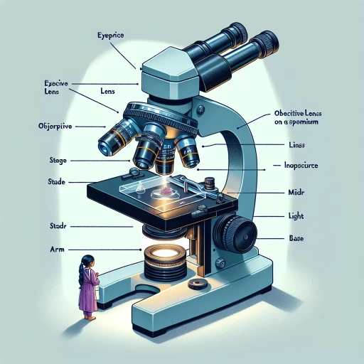
Understanding the Basic Components of a Microscope
A microscope is a complex instrument that consists of several basic components working together to magnify and resolve the details of microscopic objects. To understand how a microscope functions, it is essential to familiarize oneself with its fundamental parts. The microscope's optical system, mechanical stage, and illumination system are the three primary components that enable it to produce high-quality images. The optical system, comprising the eyepiece and objective lenses, plays a crucial role in magnifying the specimen being observed. The mechanical stage, which includes the stage and focus knobs, allows for precise movement and positioning of the specimen. Finally, the illumination system, consisting of the light source and condenser lens, provides the necessary light to illuminate the specimen. By understanding the functions of these components, one can appreciate the intricacies of microscope design and operation. The eyepiece and objective lenses are the first point of contact for light passing through the microscope, and their quality and design significantly impact the overall performance of the instrument.
The Eyepiece and Objective Lenses
The eyepiece and objective lenses are two of the most critical components of a microscope, working together to magnify and resolve the image being observed. The eyepiece lens, typically located at the top of the microscope, is responsible for further magnifying the image formed by the objective lens. It usually has a fixed magnification power, such as 10x or 15x, and is designed to work in conjunction with the objective lens to produce a clear and detailed image. The objective lens, on the other hand, is positioned near the specimen being observed and is responsible for collecting light from the sample and forming an initial image. Objective lenses come in various magnification powers, such as 4x, 10x, 40x, and 100x, and are designed to work with specific types of specimens and lighting conditions. The combination of the eyepiece and objective lenses determines the overall magnification power of the microscope, with the total magnification being the product of the two lenses' magnification powers. For example, a microscope with a 10x eyepiece lens and a 40x objective lens would have a total magnification power of 400x. The quality and type of eyepiece and objective lenses used can significantly impact the overall performance and image quality of the microscope, making them essential components for any microscopy application.
The Stage and Focus Knobs
The stage and focus knobs are two essential components of a microscope that work together to help you achieve clear and precise images of the specimen being observed. The stage is a flat platform that holds the microscope slide in place, allowing you to move it horizontally and vertically to position the specimen under the objective lens. The stage typically has clips or a mechanical stage that can be adjusted to secure the slide firmly in place. The focus knobs, on the other hand, are used to adjust the distance between the objective lens and the specimen, allowing you to bring the image into sharp focus. The coarse focus knob is used for larger adjustments, while the fine focus knob is used for more precise adjustments. By working together, the stage and focus knobs enable you to position the specimen correctly and achieve a clear and detailed image of the microscopic world.
The Light Source and Condenser Lens
The light source and condenser lens are two crucial components of a microscope that work together to provide optimal illumination for observing specimens. The light source, typically a built-in LED or halogen lamp, produces a bright, white light that is directed towards the specimen being observed. The condenser lens, located below the stage, collects and focuses this light onto the specimen, ensuring that it is evenly illuminated. The condenser lens is usually adjustable, allowing the user to control the amount of light that reaches the specimen and to optimize the contrast and resolution of the image being observed. By working together, the light source and condenser lens enable the microscope to produce high-quality images with excellent brightness, contrast, and resolution, making it possible to observe even the smallest details of the specimen being studied.
How Light Travels Through a Microscope
The journey of light through a microscope is a complex process that involves several stages, each playing a crucial role in the formation of the final image. When light travels through a microscope, it undergoes a series of transformations that allow us to observe the microscopic world in detail. The process begins with the transmission of light through the sample being observed, where the light interacts with the sample's structure and composition. As the light passes through the sample, it is refracted and magnified by the microscope's lenses, allowing us to see the sample's features in greater detail. Finally, the light is focused to form the final image, which is then observed by the user. In this article, we will delve into the specifics of how light travels through a microscope, starting with the transmission of light through the sample.
Transmission of Light Through the Sample
The transmission of light through a sample is a critical aspect of microscopy, as it allows researchers to observe the internal structures and details of the sample being studied. When light passes through a sample, it encounters various obstacles, such as cells, tissues, and other microscopic features, which can absorb, scatter, or transmit the light. The amount of light that is transmitted through the sample depends on the properties of the sample itself, including its thickness, density, and composition. Thicker samples tend to absorb more light, while denser samples may scatter light in different directions. The wavelength of the light used also plays a crucial role, as shorter wavelengths are more easily scattered by smaller particles, while longer wavelengths are more easily transmitted through the sample. To optimize the transmission of light, microscopists often use techniques such as staining, which involves applying dyes or other chemicals to the sample to enhance contrast and visibility. Additionally, the use of polarized light can help to reduce glare and improve the resolution of the image. By carefully controlling the transmission of light through the sample, researchers can gain a deeper understanding of the sample's internal structure and composition, and make more accurate observations and measurements.
Refraction and Magnification of Light
Refraction and magnification of light are two fundamental concepts that play a crucial role in the functioning of a microscope. Refraction occurs when light passes from one medium to another with a different optical density, causing the light to bend. In a microscope, refraction happens when light passes from the air into the glass lens, and then again when it passes from the glass lens into the specimen being observed. This bending of light allows the microscope to focus the light onto the specimen, creating a magnified image. The degree of bending, or refraction, depends on the angle of incidence and the refractive indices of the two media. The refractive index of a medium is a measure of how much it bends light, with higher indices indicating greater bending. In a microscope, the objective lens has a higher refractive index than the surrounding air, allowing it to bend light more efficiently and create a sharper image. The eyepiece lens also has a higher refractive index, which further magnifies the image. The combination of refraction and magnification in a microscope allows for the observation of tiny details that would be invisible to the naked eye. By carefully controlling the refraction and magnification of light, microscopes can produce high-resolution images of specimens, enabling scientists to study the microscopic world in unprecedented detail.
Formation of the Final Image
The formation of the final image in a microscope is a complex process that involves the coordination of multiple optical components. The objective lens, which is positioned closest to the specimen being observed, collects light from the sample and creates an inverted and magnified image. This image is then passed through the eyepiece lens, which further magnifies the image and corrects for any aberrations. The resulting image is then projected onto the retina of the observer's eye, where it is perceived as a clear and detailed representation of the specimen being studied. The final image is also influenced by the numerical aperture of the objective lens, which determines the resolution and contrast of the image. A higher numerical aperture allows for a greater amount of light to enter the lens, resulting in a brighter and more detailed image. Additionally, the use of a condenser lens can help to focus the light onto the specimen, increasing the intensity of the image and improving its overall quality. Overall, the formation of the final image in a microscope is a critical process that requires the precise alignment and coordination of multiple optical components.
Types of Microscopes and Their Working Principles
Microscopes are essential tools in various scientific fields, including biology, chemistry, and medicine. They enable researchers to study microorganisms, cells, and tissues in detail, leading to a deeper understanding of the microscopic world. There are several types of microscopes, each with its unique working principles and applications. Optical microscopes, for instance, use visible light to illuminate samples, while electron microscopes utilize electron beams to produce high-resolution images. Specialized microscopes, such as fluorescence and confocal microscopes, offer advanced imaging capabilities for specific research needs. In this article, we will explore the different types of microscopes, their working principles, and their applications. We will begin by examining optical microscopes and their limitations, which will provide a foundation for understanding the advantages and applications of other types of microscopes.
Optical Microscopes and Their Limitations
The optical microscope, also known as a light microscope, is a type of microscope that uses visible light and a system of lenses to magnify images of small objects being observed. Optical microscopes are the most common type of microscope and are widely used in various fields such as biology, medicine, and materials science. They work by using a light source to illuminate the sample being observed, and then using a combination of objective lenses and eyepiece lenses to magnify the image. The objective lenses collect light from the sample and create an image, which is then magnified by the eyepiece lenses. Optical microscopes can produce high-quality images with good resolution, but they have some limitations. One of the main limitations of optical microscopes is their resolution, which is limited by the wavelength of light used. The resolution of an optical microscope is typically around 200-300 nanometers, which means that it cannot resolve objects that are smaller than this size. Another limitation of optical microscopes is their depth of field, which is the range of distances within which the image is in focus. Optical microscopes have a relatively shallow depth of field, which means that only a small portion of the sample is in focus at any given time. This can make it difficult to observe samples that have a lot of depth or complexity. Additionally, optical microscopes can be affected by aberrations, which are distortions in the image caused by the lenses. Aberrations can reduce the quality of the image and make it more difficult to interpret. Overall, optical microscopes are powerful tools for observing small objects, but they have some limitations that need to be considered when using them.
Electron Microscopes and Their Advantages
Electron microscopes are a type of microscope that uses a beam of electrons to produce an image of a sample being observed. They have several advantages over traditional light microscopes, including higher resolution and magnification, allowing for the observation of smaller structures and details. Electron microscopes can achieve resolutions as low as 0.1 nanometers, compared to light microscopes which are limited to around 200 nanometers. This makes them ideal for studying the fine details of cells, viruses, and other small biological structures. Additionally, electron microscopes can be used to study the surface morphology of materials, making them useful in fields such as materials science and nanotechnology. Another advantage of electron microscopes is their ability to produce three-dimensional images, allowing researchers to gain a better understanding of the structure and organization of complex biological systems. Overall, electron microscopes offer a powerful tool for scientists and researchers, enabling them to study the microscopic world in greater detail than ever before.
Specialized Microscopes and Their Applications
Specialized microscopes have revolutionized various fields of science, medicine, and research by providing high-resolution imaging and analysis capabilities. One such microscope is the Electron Microscope (EM), which uses a beam of electrons to produce high-resolution images of nanoscale structures. The EM is widely used in materials science, biology, and medicine to study the morphology of cells, tissues, and materials at the nanoscale. Another specialized microscope is the Atomic Force Microscope (AFM), which uses a physical probe to scan the surface of a sample, providing high-resolution images of surface topography and mechanical properties. The AFM is commonly used in materials science, nanotechnology, and biophysics to study the properties of surfaces and interfaces. The Scanning Tunneling Microscope (STM) is another type of specialized microscope that uses a sharp probe to scan the surface of a sample, providing high-resolution images of surface topography and electronic properties. The STM is widely used in materials science, nanotechnology, and physics to study the properties of surfaces and interfaces at the atomic scale. The Confocal Microscope is a specialized microscope that uses a laser to excite fluorescent dyes in a sample, providing high-resolution images of cellular structures and dynamics. The Confocal Microscope is widely used in biology, medicine, and neuroscience to study the behavior of cells and tissues in real-time. The Super-Resolution Microscope is a specialized microscope that uses advanced imaging techniques to produce high-resolution images of cellular structures and dynamics at the nanoscale. The Super-Resolution Microscope is widely used in biology, medicine, and neuroscience to study the behavior of cells and tissues at the nanoscale. These specialized microscopes have greatly advanced our understanding of the structure and function of cells, tissues, and materials, and have opened up new avenues for research and discovery in various fields of science and medicine.