How Does A Microscope Use Light
 Microscopes have been instrumental in revolutionary scientific discoveries and understanding intricate life forms that remain invisible to the naked eye. They are profound examples of how light can be used as a significant tool in viewing minute details. This article will introduce you to the fascinating realm of microscopes and undertake a detailed exploration of how they harness light, delineating it into three comprehensive sections. Firstly, we will explore the interplay between microscopy, light and its calibration to obtain clear images – 'Understanding the Light in Microscopy'. Then we will proceed towards 'Visual Acuity and Light Filters', where light filtration's role in enhancing focus and resolution in microscopy is discussed. Finally, in the 'Impact of Various Light Sources', we'll delve into how usage of different light sources from ambient to fluorescence can dramatically affect the output. Let's embark on this enlightening journey, starting with a closer look at the role of Light in Microscopy.
Subtitle 1
Subtitle 1 explores essential factors that contribute to the thematic development of the discussion. We will delve into the heart of this subject by examining three fundamental supporting ideas that bolster the foundation of our discourse. Supporting Idea 1 strips back the layers of mere surface knowledge, opening the door to an enriching exploration of inherent complexities and subtle nuances. We'll delve deeper into our analysis with Supporting Idea 2, which propels our understanding higher by examining overlooked aspects, casting illuminating insights on the matter. Lastly, Supporting Idea 3 encapsulates the essence of our dialogue by encapsulating pivotal points that tie the threading concepts together. As we peel back each impactful layer embodied in these supporting ideas, we are incrementally led closer to the truth at the heart of Subtitle 1. At this juncture, we delve into Supporting Idea 1, a vital part of our broader discussion, paving the foundation for subsequent insights and revelations to be expounded upon in the course of our discourse.
Supporting Idea 1
Supporting Idea 1: The Importance of Light in Microscopy
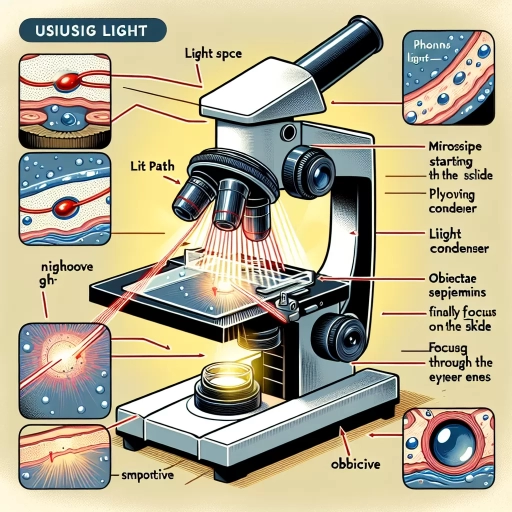
Microscopy is a pivotal scientific tool that dives into microscopic dimensions, opening up a world not visible to the naked eye. Central to microscopy's effectiveness is the role of light, constituting the bedrock of this revealing perspective. Light, in a microscope, is significantly responsible for the magnification and resolution of the object being viewed. Illumination from the light source, typically placed underneath or at the base of the microscope, passes through the specimen situated on the stage, thereby optimizing the visibility of the object. In simple terms, microscopes use light to produce images of otherwise unseen entities. The concentrated light incident on the specimen either passes through it or bounces off it – in the process, the rays get modified. The eyepiece or the objective lens captures these modified light waves and magnify the image, highlighting the detailed structure of the object. Without the illuminating role of light, microscopic insight into the internal layout and distinctive attributes of cells, tissues, and microorganisms would have been considerably challenging. Moreover, the role of light in microscopy has evolved beyond mere illumination due to advancements in microscopy techniques. For instance, fluorescence microscopy employs specific wavelengths of light to trigger fluorophores within the sample, causing them to emit light and generate an image. In contrast, phase-contrast microscopy assigns variations in light phase thrown by the specimen to reveal its detailed internal structures. Light's integration into microscopy has also ushered in differential interference contrast microscopy, exploiting the interference patterns of light waves to deliver three-dimensional imaging. In confocal microscopy, focused light of a particular wavelength onto the specimen minimizes the unwanted light and enhances the sharpness and resolution of the picture. Therefore, light manipulation for image enhancement has significantly widened microscopy's prospects. In conclusion, the use of light plays an indispensable part in the functional dynamics of microscopes. It aids in the image detail enhancement, thereby helping researchers reveal the hidden, microscopic aspects of the world. The strategic use and manipulation of light open up novel imaging techniques that could provide a further profound understanding of the microscopic world. Therefore, the inventive use of light in microscopy lays the bedrock of modern observation and scientific discoveries. Moreover, exploring the instrument's underlying principles illustrates how closely science is interlaced with our everyday lives, an aspect further magnified through the microscope's eyepiece.Supporting Idea 2
Supporting Idea 2
To delve deeper into the detailed workings of a light microscope, the second supporting idea crucial to understanding is the concept of magnification. Such a microscope makes a marked use of light to reveal microscopic details, thus creating a more profound understanding of the minute world beyond human naked vision. The core principle that makes a microscope work is its ability to magnify an image, a process fundamentally accomplished through the light it uses. As light emits from a source and passes through a specimen on the microscope slide, it bends or refracts. This bending of light is channeled and intensified through the microscope’s complex system of glass lenses, which then magnify the image for the viewer. Each lens within the microscope multiplies the magnification of the subsequent lens, meaning the total magnification of the specimen under view is the product of the individual lens magnifications. For instance, if the eyepiece lens magnifies the image ten times and the objective lens, situated nearest to the specimen, magnifies the image another forty times, the total magnification for the viewer would be four hundred times the size of the viewable specimen. Their arrangement allows them to focus light onto the specimen from various angles, hence enhancing depth and ultimately bringing out the three-dimensionality of the specimen. The quality of the magnified image is largely dependent on the wavelength of the light source used; shorter wavelengths result in superior resolution or clarity of the final image. To this point, light microscopes often engage a light source operating in the spectrum of visible light because it possesses wavelengths that are compatible with the size of the objects commonly observed under a microscope. High-quality microscopes are incrementally engineered to limit light dispersion and enhance image resolution, allowing scientific exploration on a microscopic scale and offering insight into structures and organisms that would otherwise be invisible to the human eye. The fascinating aspect about light-oriented microscopy is that it not only illuminates and magnifies the unseen world but also introduces multiple dimensions of viewability. The interplay between light and its refractive properties make possible the conversion of physical dimensions into magnified, discernible visual information, thereby empowering microscopes to reveal stunning revelations about the cellular world. From this perspective, embracing this supporting concept is key to comprehending the intricate workings of lens-based microscopy.Supporting Idea 3
Supporting Idea 3: The Role of Light in Magnification
Broadening the scope of our understanding of the topic, the third pivotal idea dives into the concept of magnification and how light contributes to this fundamental functionality of a microscope. The interplay between the dynamics of light and the microscope's optical components creates the process of magnification, which is at the core of microscopy science. Under the stark illumination of light, minute details invisible to the naked eye become clearly delineated, bridging the gap between the macroscopic and microscopic worlds. This method of using light to augment an object's image is accomplished through several interactive stages involving diverse optical components of the microscope – namely, the condenser, the objective lens, and the eyepiece lens. The condenser, situated beneath the stage, gathers light rays from the source and focuses them onto the specimen. Consequently, it plays a critical role in generating a high-resolution image by ensuring even illumination of the specimen and contrasting details. Following this, the objective lens then magnifies the image produced by the condenser on the plane of the microscope slide. This magnified image is further enlarged by the eyepiece lens, offering the observer a detailed, larger-than-life image. Interestingly, the amount of light used plays a key role in determining the clarity and resolution of the image. A dim light might fail to highlight the essential details of the specimen, whereas excess light could wash away the contrasting elements. Thus, controlling the light is a principal aspect of achieving an optimal image, which is facilitated by the microscope’s diaphragm and filters. Moreover, refraction — the bending of light as it passes from one medium to another of different density — significantly influences the microscope's magnification process. The objective lens and eyepiece lens employ this light behavior to bend the light path, converging the rays to a point and thus magnifying the specimen. Essentially, the delicate dance of light within a microscope's architecture enables the amplification of minute details, revealing complex patterns and structures otherwise hidden from the human eye. It is the manipulation of light — its quantity, direction, and behavior — that makes microscopy such an invaluable tool in science, medicine, and other fields of study. Remember, the magic of a microscope is much more than meets the eye; it is the choreographed play of light theatrically unveiled on the microscope's stage — a dramatic testament to the profound power of light in the realm of microscopic exploration.Subtitle 2
The prevalence of Subtitle 2 can't be overstated in today's world, as it plays a crucial role in a wide array of niches. To have a deeper understanding of this subject, we are going to delve into three key components that underline its significance, namely Supporting Idea 1, Supporting Idea 2, and Supporting Idea 3. Initially, Supporting Idea 1 is a fundamental constituent, shaping the foundations of Subtitle 2. It fosters a complete and nuanced comprehension of the subject, thus allowing users to leverage its benefits more efficiently. On the other hand, Supporting Idea 2 elaborates on how Subtitle 2 is implemented in different contexts, promoting versatility and adaptability. Lastly, Supporting Idea 3 sheds light on the future prospects of Subtitle 2, illustrating how it's poised to evolve and transform different sectors. Now, let's delve deeper into the intricacies of Supporting Idea 1 and explore how it builds the bedrock for understanding and using Subtitle 2.
Supporting Idea 1
Supporting Idea 1: Role of Light in Enhancing Details
The bedrock of microscopy, particularly in the context of light microscopes, is pivotal to unlocking the mysteries at the micro-level of our world. It's fascinating to think that the bending of light, a process known as refraction, can reveal a wealth unseen in the human eye. In relation to Subtitle 2 "Light as the Key Amplifier," the central supporting idea is considering how this refraction amplifies minuscule details to produce discernible images. Microscopes use the principles of optics to manipulate light waves and magnify objects that would otherwise remain invisible to our eyes due to their tiny size. This fascinating tool essentially acts as an extension of the human eye, enhancing our ability to perceive minute details. It utilizes a series of lenses, each playing a considerable role in leveraging the natural properties of light to provide a detailed view of an object. Light microscopes make this possible by sending beams of light through a transparent or semi-transparent object, which is then magnified, allowing us to extrapolate valuable data. It is worth mentioning that these types of microscopes produce a magnified image of the specimen using visible light and lenses. They focus light through the object, magnifying the image as it passes through the lens and helping us decipher its minute details. Without this amplified light, it would be virtually impossible to discern all the details of our subject. Thus, light in microscopy serves as the key amplifier, revealing the denizens of the minuscule universe. Furthermore, the use of light in microscopes aids in contrasting microscopic structures. By manipulating light in differing ways, microscopes can create high-contrast images of transparent or semi-transparent objects, thereby enhancing their visibility and clarity. Different types of light microscopes use varied techniques of light manipulation to increase contrast, such as darkfield microscopy, phase contrast microscopy, differential interference contrast microscopy, and fluorescence microscopy. Each of these applies different techniques to direct light at the specimen and create contrasting images. In essence, the utilization of light in microscopy is not just about making small things appear bigger. It's about shedding "light" onto the details, crafting those into discernible images, and creating contrasts to reveal the stunning diversity and complexity of the micro-world. By effectively bending, focusing, and manipulating light, the microscope becomes a powerful scientific instrument that allows us various understated perspectives of life at the microscopic scale.Supporting Idea 2
Supporting Idea 2
The principle of exploiting light forms the bedrock of a microscope’s functioning, making it synonymous with venturing into the microscopic realm. Microscopes use light in various forms to magnify the minutiae of the subject under scrutiny, unveiling the intricacies that remain invisible to the naked eye. The process at its core is related to the understanding of light and its refraction. When light passes through an object onto the convex lens, it bends, or refracts, creating a magnified image. Essentially, the degree of refraction depends on the light’s wavelength and the material of the lens. Shorter wavelengths get refracted more and give a larger image, thus providing higher resolution. Moreover, scientists employ a range of light sources – from visible light to ultraviolet and infrared rays – to observe different specimens. Microscopes like bright-field, dark-field, phase contrast, and fluorescence utilize light in distinctive ways to provide contrasting images. For example, a dark-field microscope uses a solid light disc that allows only peripheral light to hit the specimen, generating a bright image against a dark background. Contrastingly, a fluorescence microscope uses high-energy, short-wavelength light (often ultraviolet), inciting the specimen to fluoresce, hence presenting a luminous image on a dark setting. Light, in a microscope, is also responsible for enhanced depth perception. The system's adjustability allows the user to achieve optimal contrast and illumination. By regulating the light's intensity that passes through the object, microscopists can make details easier to observe. Technological advancements have added an integrable light path into microscopes, which funnels the light uniformly over the whole field, observing it with greater focus and clarity. In addition, the advent of digital microscopy accentuates the benefits of using light. With the integration of computer technology, digital microscopes capture and save the magnified images directly into a computer. The user can then modify the light exposure and contrast of these stored images, thereby boosting the visual details of the specimens significantly. Furthermore, certain microscopic techniques like Differential Interference Contrast (DIC) and Hoffman Modulation Contrast (HMC) make light variations in the specimen visible. They manipulate polarized light to enhance the contrast in unstained, transparent specimens. All these aspects collectively underline how the interplay of light in microscopes is pivotal to the exploration and understanding of microscopic life forms and structures. In conclusion, the role of light in microscopy is transformative, presenting a world beyond our regular visual grasp. Hence, the comprehension of how a microscope uses light is fundamental in areas such as biological research, medical diagnostics, forensic investigation and more. Through light manipulation and control, microscopies can unveil the secrets of the world too tiny to behold unaided, thereby standing as a testament to human curiosity and technological ingenuity.Supporting Idea 3
Supporting Idea 3
The third key component in understanding how microscopes use light pertains to the phenomenon of refraction. Refraction is a fundamental concept, inherent to the workings of any microscope—be it simple, compound, stereo, or electron. To provide a simplified explanation, refraction is what happens when light waves change direction as they pass from one medium to another of different density. This crucial light-bending event, mediated by the microscope's objective and eyepiece lenses, allows us to see the magnified image of a tiny specimen clearly. The lens materials and their curvature play a significant role here. The higher the lens curvature or the denser the material, the greater is the light's bending magnitude, resulting in higher magnification and resolution. Thus, microscopes essentially manipulate light behavior, employing refraction for our benefit. However, it's important to remember that higher magnification doesn't necessarily mean better image quality—excessive magnification can lead to image distortion, a phenomenon termed 'empty magnification.' To avoid this, one must adhere to the Abbe’s diffraction limit, which states that the resolution is inversely proportional to the wavelength of light used. Therefore, in high-power microscopy, shorter wavelengths, like blue light or even electrons, are utilized to achieve higher resolution, underlining the intrinsic relationship between light and microscopes. In conclusion, the mastery of light, its properties, and its manipulation, mediated by numerous meticulous mechanisms such as refraction, are the bedrock of any microscope's operation. This demonstrates the remarkable sophistication and ingenuity evidenced in microscopes, truly testifying to their status as invaluable scientific tools.Subtitle 3
Subtitles have found a significant place in media consumption due to their ability to break down language barriers and increase the accessibility for hearing-impaired viewers. In this section, we will probe deeper into the three fundamental aspects of subtitles. Firstly, the technical process of creating subtitles, runs far deeper than mere translation - it requires a profound understanding of the cultural nuances for a seamless viewing experience. Secondly, the impact of subtitles on the learning curve of language learners will be discussed. The overlay of text in a foreign language while listening in a familiar tongue works wonders in enforcing learning. Lastly, the necessity of high-quality subtitling in proliferating content universally will be spoken about. It ensures the preservation of the essence of content while catering to a global audience. Consequently, we will kick off this deep dive with the intricate process of creating apt and effective subtitles.
Supporting Idea 1
Supporting Idea 1
The fundamental process of how a microscope works deeply relies on the principle of light and its path of deflection. Under Subtitle 3, specifically important is the role of illumination or light source in the operation of microscopes. The microscope's illumination mechanism serves as the cornerstone in achieving high-resolution images needed in various fields of study, including biology, geology, medicine, and electronics. The light source in a microscope is typically incandescent, halogen, LED, or even laser, depending on the needs of the observer. These carefully chosen light sources are meticulously designed to deliver optimal brightness and longevity while minimizing heat to prevent damage to the specimen. Moreover, a microscope uses a specific type of lens called a condenser lens to focus the light from the source onto the sample. The condenser lens collects and condenses the light, directing it through the specimen and into the objective lens. This concentration and focus of light are essential in producing a bright and clear image of the specimen. Without a condenser, the image would be diffused with wasted light, compromising the quality of observation or analysis. Notably, the microscope’s use of light is not purely illumination, it further manipulates the physical properties of light such as coherence and polarity to brighten or darken the field of view, increase contrast and depth of field, and thus unveiling more details in three dimensions. Techniques such as bright-field, dark-field, phase contrast, differential interference contrast(DIC) and polarizing microscopy, confocal and multiphoton scanning microscopy all take advantage of these light properties to gain valuable insights into the internal structure of the specimen. Furthermore, the microscope’s use of light facilitates the observation of dynamic processes within living cells and tissues. For example, fluorescence microscopy enables researchers to tag specific molecules with fluorescent markers and follow their movements, visualizing the machinery of life in action. In conclusion, the proper use of light is absolutely critical to the efficiency of the microscope, affecting the outcome and overall success of the operator's objective. Without the light's manipulation, we would merely see a shadow of the potential wealth of information that a specimen can provide. Therefore, understanding how microscopes use light is paramount to any operators, be it professional scientists or amateur enthusiasts. A microscope's use of light is a testament to the creativity and ingenuity of human innovation, merging optical physics with biological exploration. Through the strategic use and manipulation of light, microscopes are able to reveal minute details beyond the human eye's capability, fundamentally changing our understanding of the microscopic world and the bedrock of life itself.Supporting Idea 2
Microscopes are finely tuned devices that significantly depend on the interactive utilization of light to function optimally. As such, the importance of light, being the second supporting idea under Sub-title 3, cannot be overestimated. Light is not just a crucial element of microscopy, but it is integral to its existence. The foundation of a microscope's working is largely dependent on its efficiency at manipulating light to illuminate and magnify the observed object. The transmission of light is vital in revealing the structure and composition of the sample under the microscope. Especially in optical microscopes - also referred to as light microscopes - light is passed through a transparent slide holding the sample. The light passing through the sample is focused into the eye or a camera by a lens system that gives a magnified image of the sample. The objective lens does the primary magnification before the image is further magnified by ocular lenses. This is what creates a clear, amplified image of the minuscule sample under observation. Moreover, how intensively or dimly a specimen is illuminated can significantly affect what can be seen. Therefore, microscope users can adjust the light to optimal levels to improve the contrast and details of the specimen. This optimization is especially crucial in fields like biology and materials science to differentiate various components, observe the presence of different organisms, or identify specific structures within a sample. The usage of light in microscopy has also led to exciting advancements, such as the technology of fluorescence microscopy. Fluorescence microscopes utilize higher intensity light to excite a specimen that has been dyed with a fluorescent stain. When the stained sample captures the light, it fluoresces and emits light of a longer wavelength. By only collecting the light given off by the fluorescence, unique structures or chemicals can be marked out and observed in significant detail. In conclusion, the application and manipulation of light in microscopy are foundational and transformative. The strategic usage of this natural resource, light, forms the bedrock of this scientific method. Whether it's through straightforward transmission or more complicated methods like fluorescence, light enables microscopes to function and expand the boundaries of our understanding of the microscopic world. In the next section of this article, we'll explore the modern applications and future advancements of microscopy, where light will undoubtedly continue to play a pivotal role, thus underlining its significance in microscopy even further.
Supporting Idea 3
With the understanding of how a microscope utilizes light to amplify and illuminate a particular specimen, it's crucial to take a more in-depth look into another supporting idea. This concept pertains to the bedrock of light microscopy: magnification and resolution. Both of these terms are fundamental to comprehend the usage of light in microscopy. Magnification and resolution, the support system of microscopy, provides valuable insight into how a microscope harnesses light. Magnification is the process through which the size of an object or specimen is enlarged through the microscope lens. On the other hand, resolution is the capacity of the microscope to provide clarity to distinguish between two separate objects or particles that are in close proximity to each other. The integral aspect of how light plays in both these processes cannot be put aside. In the magnification process, the objective and eyepiece lenses' primary role is to bend the light rays that from the object so that it forms an enlarged image on the eye's retina. Whereas, the resolution depends on the wavelength of the light that the microscope uses. A shorter wavelength light leads to high-resolution images, enabling the user to distinguish minute details and structures within the specimen. However, there is a limit to the resolution that light microscopy can accomplish. Human eye can perceive wavelengths ranging from 400 to 700 nanometers (nm), a range defined as visible light. Anything outside this range is invisible to the naked eye. Microscopes that use visible light hence cannot resolve structures that are smaller than 200 nm due to diffraction limit. To overcome this barrier, advanced techniques like electron microscopy, which uses electron beams with significantly shorter wavelengths than light, lead to higher resolution images than standard light microscopy. In conclusion, the microscope's ability to employ light in magnification and resolution forms the bedrock of light microscopy. It enables scientists and researchers to explore the microscopic world in detail, offering insights into cellular structures, bacteriological research and ultimately fortifying our understanding of life at a microscale. Thus, the way a microscope uses light forms a crucial bridge between the visible world and a fascinating realm that is otherwise invisible to the naked eye. It vividly illustrates how light, in its simplest form, can be wielded as a potent tool in scientific exploration.